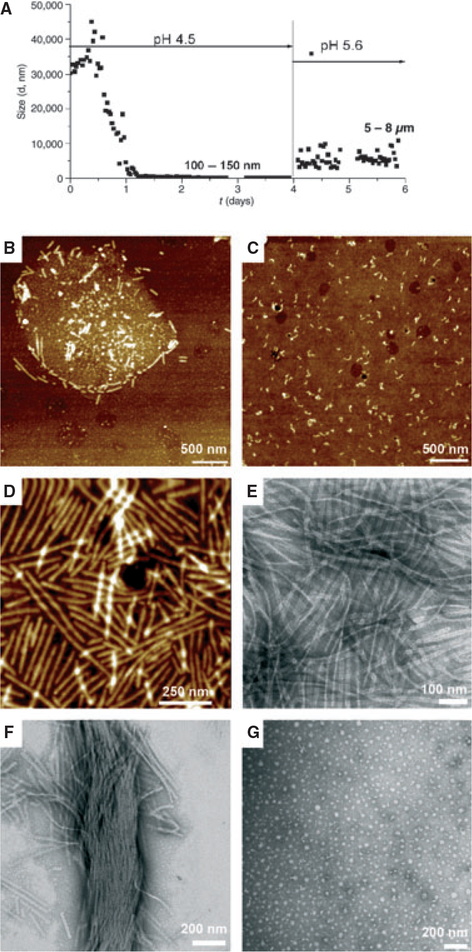
Fig. 1.
Amelogenin rH174: time-course of assembly and ribbon elongation at the water–oil interface as a function of pH and the water–oil ratio. Metastable water-in-oil emulsions of amelogenin (0.37 mg ml−1) were prepared by mixing with octanol/ethyl acetate (water/oil ratio = 1:4) in the presence of high concentrations of calcium and phosphate ions at pH 4.5 [degree of saturation of hydroxyapatite (DSHA) = 3.7]. (A) Particle size analysis by time-resolved dynamic light scattering. The particle size decreased over time from micrometer-size to nanometer-size particles of 100–150 nm. At day 4, the pH was increased to 5.6 (DSHA = 11–12) and the particle size increased to 5–8 μm. Atomic force microscopy topographic images (tapping mode) and transmission electron microscopy images of supramolecular amelogenin microstructures and nanostructures were obtained. Micelle structures and ribbon-like structures were observed within the first minute of emulsion preparation and immobilization on a glass substrate (B); short ribbons appeared in the water phase after 4 d (C). Abundant elongated amelogenin nanoribbons (D, E) and bundles (F) formed at pH 5.6. In the absence of calcium, amelogenin assembled into characteristic 20-nm nanospheres (G).

