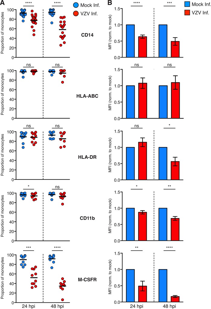
FIG 7.
Cell surface immunophenotyping of VZV-infected monocytes. (A) Mock-infected monocytes (blue) and VZV-infected (red) monocytes were examined for the proportions of cells expressing the indicated cell surface markers at 24 and 48 hpi by flow cytometry. Gating was performed on live, single cells, prior to assessment of VZV infection by VZV gE:gI staining. Symbols depict the proportions of monocytes from multiple independent donors expressing the indicated markers, and bars represent the mean. (B) The MFI of immune markers on VZV-infected monocytes was normalized to that of the respective mock-infected monocytes at each time point. Bars represent the mean MFI (±SEM). Results are from multiple independent experiments examining CD11b (n = 8), HLA-ABC (n ≥ 8), HLA-DR (n ≥ 8), CD14 (n ≥ 18), and M-CSFR (n ≥ 9). Statistical analyses were performed for proportional and MFI analysis by paired two-tailed Student's t test. *, P < 0.05; **, P < 0.005; ***, P < 0.0005; ****, P < 0.00005; ns, not significant.

