FIG 8.

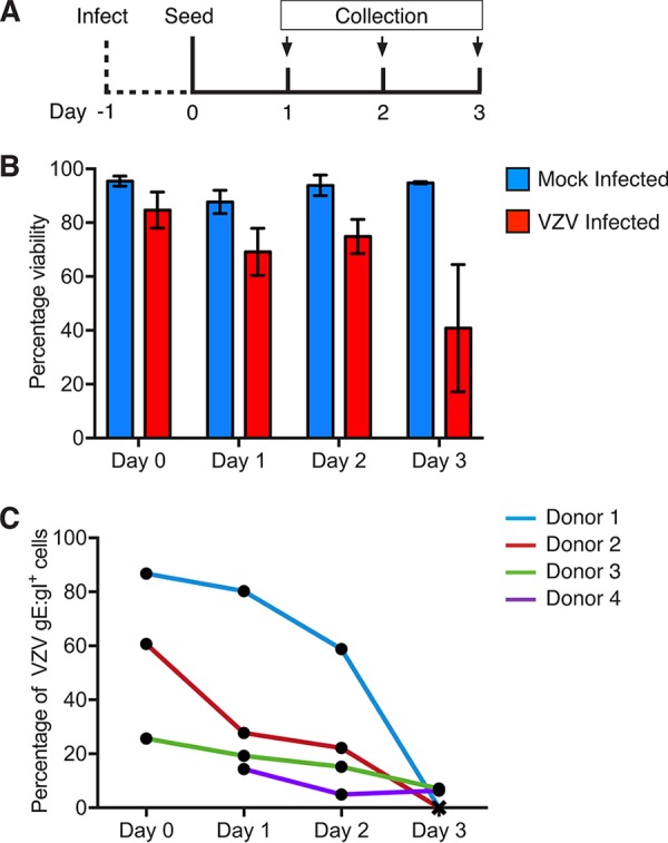
Differentiation of VZV-infected monocytes. Mock- and VZV-infected monocytes were collected at 24 hpi (day 0) and adhered into tissue culture plates in serum-free medium for 2 h at 37°C under 5% CO2. Nonadherent monocytes were aspirated, and the cultures were maintained for 3 days in medium supplemented with 25 ng/ml M-CSF (Peprotech). Adherent monocytes were collected by gentle scraping into the medium, stained for viability and VZV gE:gI, and analyzed by flow cytometry. (A) Timeline for infection and seeding. (B) Proportions of viable adherent monocytes cultured with mock-infected (blue) or VZV-infected (red) HFF. (C) Proportions of VZV gE:gI+ monocytes across each day measured for four independent donors.
