FIG 9.

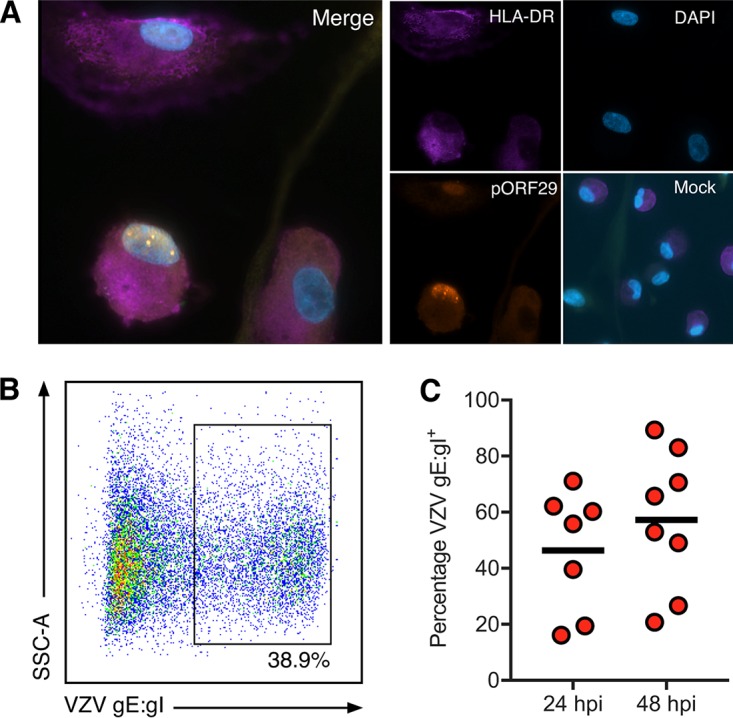
VZV infection of monocyte-derived macrophages. Freshly isolated monocytes were adhered under serum-free conditions as described in the text and cultured in 25 ng/ml M-CSF for 6 days to generate macrophages (M-Mφ). M-CSF macrophages were cocultured with VZV-infected CFSE-labeled HFF for 24 and 48 h. VZV exposed M-Mφ were examined by IFA for the presence of HLA-DR and VZV pOR29. (A) Merged IFA image of VZV-infected M-Mφ at 48 hpi. Individual channels of HLA-DR (purple), pORF29 (orange), and DAPI (blue) are shown. A merged image of mock-infected M-Mφ at the same time point is also shown. All images were taken at a ×20 magnification and are representative of those from 4 independent experiments. Macrophages were collected at 24 and 48 hpi and assessed for VZV gE:gI by flow cytometry. Gating was performed on live, single cells and excluded the CFSE-labeled HFF inoculum. (B) VZV-infected M-Mφ exhibited cell surface VZV gE:gI. (C) VZV-infected M-Mφ displayed surface VZV gE:gI at 24 hpi (n = 4) and 48 hpi (n = 5). Bars indicate the mean proportion at each time point.
