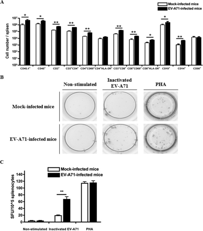
FIG 6.
Activation of human T cells and the presence of EV-A71-specific T cell responses in the spleen of humanized mice after EV-A71 infection. Four-week-old humanized mice were inoculated i.p. with or without EV-A71 (108 PFU/mouse). (A) Mononuclear cells (MNC) were isolated from spleen at 9 weeks postinfection for FACS analysis. The total number of different immune cell types in the spleen of the mock-infected (n = 4) and EV-A71-infected humanized mice (n = 4) is indicated in the graph. Data are expressed as means ± SEM. *, P < 0.05; **, P < 0.01. (B and C) MNC from mock-infected (n = 4) and EV-A71-infected humanized mice (n = 4) were isolated from the spleen at 9 weeks postinfection, and 104 MNC were stimulated with inactivated EV-A71 for 72 h for human IFN-γ ELISPOT assay. Representative ELISPOT results for nonstimulated and inactivated EV-A71-stimulated MNC from mock-infected and EV-A71-infected humanized mice are shown. PHA-stimulated cells were used as a positive control. The human IFN-γ T cell responses from each group are shown as spot-forming units (SFU) per 104 MNC. Data are expressed as means ± SEM. **, P < 0.01.

