Figure 4.
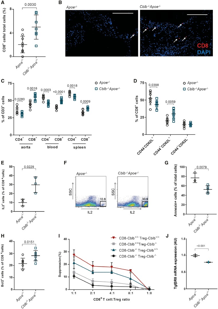
Casitas B-cell lymphoma-B deficiency increases CD8+ T cell abundance in Cblb−/−Apoe−/− mice by reducing apoptosis and regulatory T cell-mediated suppression. (A) Percentages of CD8+ T cells in advanced atherosclerotic plaques of aortic roots of 20-week-old Apoe−/− (n = 10) and Cblb−/−Apoe−/− mice (n = 7). (B) Representative pictures of anti-CD8 (Alexa Fluor 594, red) staining (DAPI staining: blue). White arrows indicate CD8+ T cells. Scale bar: 100 μm. (C) Flow cytometric analysis of CD4+ and CD8+ T cells in aortic arch, blood, and spleen of Apoe−/− and Cblb−/−Apoe−/− mice (n = 7) (D) Quantification of naïve (CD44−CD62L+), central memory (CD44+CD62L+), and effector memory (CD44+CD62L−) CD8+ T cells in spleens of Apoe−/− and Cblb−/−Apoe−/− mice. (E, F) interleukin-2 production by CD8+ T cells isolated from in vitro-restimulated splenocytes (n = 3), Representative dot plots are shown. (G) Fraction of apoptotic (Annexin V+) cells of CD3/CD28-activated isolated splenic CD8+ T cells from Apoe−/− (n = 3) or Cblb−/−Apoe−/− (n = 5) mice that were incubated with TNF for 96 h. (H) Flow cytometric analysis of BCL2 expression in CD8+ T cells (n = 7). (I) Regulatory T cell suppression assay using splenic CD8+ T cells and CD4+CD25+ regulatory T cells from Apoe−/− and Cblb−/−Apoe−/− mice, co-cultured at various ratios (n = 3 experiments). (J) TGFβRII mRNA expression in CD8+ T-cells isolated from Apoe−/− and Cblb−/−Apoe−/− mice (n = 3). Data are presented as mean ± standard deviation.

