Figure 7.
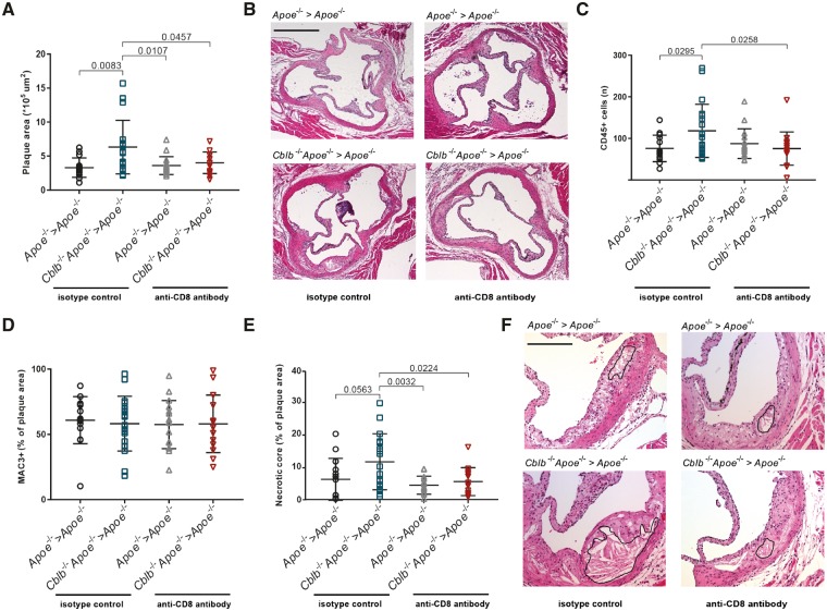
The progression of atherosclerosis is hampered in CD8+ T cell-depleted haematopoietic Cbl-b−/−Apoe−/− chimeras. (A) Aortic roots of 20-week-old haematopoietic Apoe−/− and Cblb−/−Apoe−/− chimeras analysed for the amount of atherosclerosis. (B) Representative pictures of haematoxylin and eosin-stained aortic root cross-sections containing advanced atherosclerotic plaques. Scale bar: 500 μm. (C–E) Quantification of plaque CD45+ cells, MAC3+ cells and necrotic core area in plaques of the aortic roots. Representative pictures are shown. The black line indicates the necrotic core. Scale bar: 200 μm. Data are presented as mean ± standard deviation (n = 14–18).

