
Fig. 5.
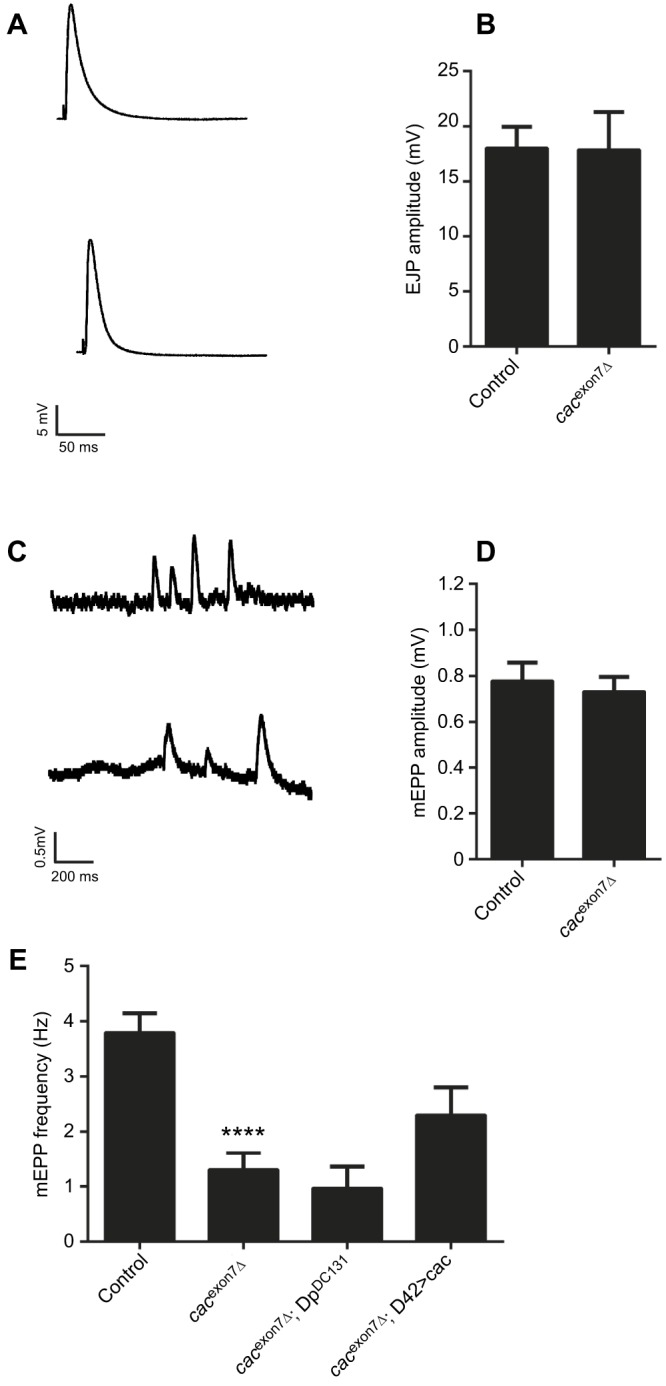
Synaptic physiology at the larval NMJ in the cacexon7Δ mutant. (A) Representative examples of excitatory junctional potentials (EJPs) from control (top) and cacexon7Δ mutant (bottom) larvae. (B) EJP amplitude was unchanged in cacexon7Δ mutants (N=11) compared to controls (N=17) (two-tailed t-test, t=0.04 d.f.=26; P>0.05). (C) Representative example of miniature end plate potentials (mEPPs) in control (top) and cacexon7Δ mutant (bottom) larvae. (D) The mEPP amplitude was unchanged in the cacexon7Δ mutant (N=11) compared to controls (N=17) (two-tailed t-test, t=0.04 d.f.=26; P>0.05). (E) The mEPP frequency was significantly reduced in the cacexon7Δ mutant (N=11) compared with controls (N=17) and was not rescued by either the DpDC131 duplication (N=5) or by expressing cacophony in motor neurons using the D42 GAL4 driver (N=9) (one-way ANOVA followed by Dunnett's multiple comparisons test, F3,38=11.2; ****P<0.0001). All experiments were carried out in 1 mmol l−1 calcium using male larvae.