
Fig. 1.
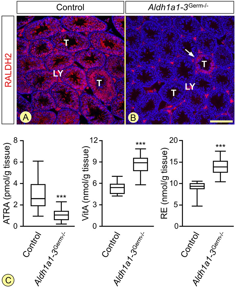
The Aldh1a1-3Germ−/− mutation suppresses RALDH2 expression in germ cells and alters testicular retinoid metabolism. (A,B) Immunohistochemical detection of RALDH2 (red signal) on histological sections from 8-week-old control and Aldh1a1-3Germ−/− mice. The white arrow in B indicates the only seminiferous tubule (T) cross-section, out of the 230, that still expresses RALDH2. The signal observed in Leydig cells (LY) is unspecific, as RALDH2 transcripts are not detected in these cells (Vernet et al., 2006b). Nuclei were counterstained with DAPI (blue signal). Scale bar: 160 µm. (C) Box-and-whisker plots showing the testicular concentrations of ATRA, vitA and retinyl esters (RE) in 9- to 10-week-old Aldh1a1-3Germ−/− (n=16) and control (n=14) mice. The boxes indicate the upper and lower quartiles, the lines inside mark the medians, and the whiskers delineate the highest and lowest values. ***P<0.001, Student's t-test.