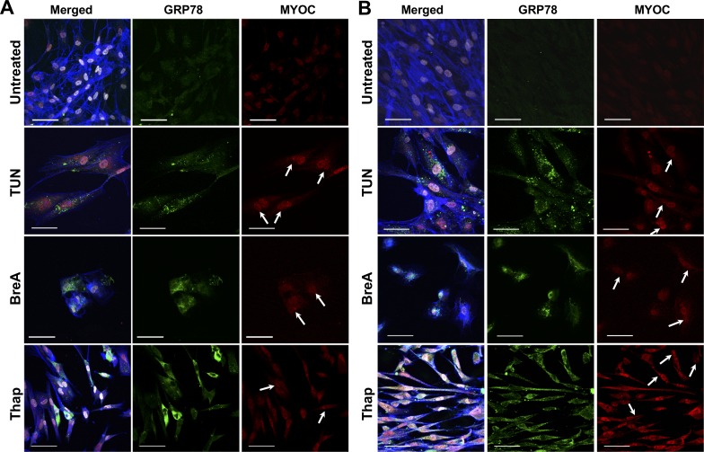
Figure 2.
Expression of GRP78 and myocilin increased after 72-hour ER stress induction. Representative immunostaining images show GRP78 (green), myocilin (MYOC, red) merged with DAPI (white), and F-actin (blue) on TMSCs (A) and TM cells (B). Myocilin (red, arrows) accumulated in the perinuclear region where the ERs are and partially overlapped with GRP78 (green). Tun and BreA reduced attached live cell numbers. Thap treatment made both TMSCs and TM cells more elongated. Scale bars: 50 μm.

