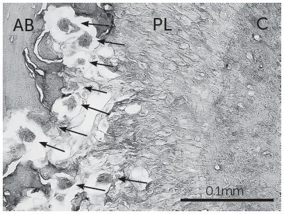
Figure 3. Micrograph of the mesial root of a first molar and its supporting tissue processed for TRAP histochemistry to identify osteoclast cells (arrows) (400x magnification). AB = alveolar bone; PL = periodontal ligament; C = cementum; bar: 0.1 mm.