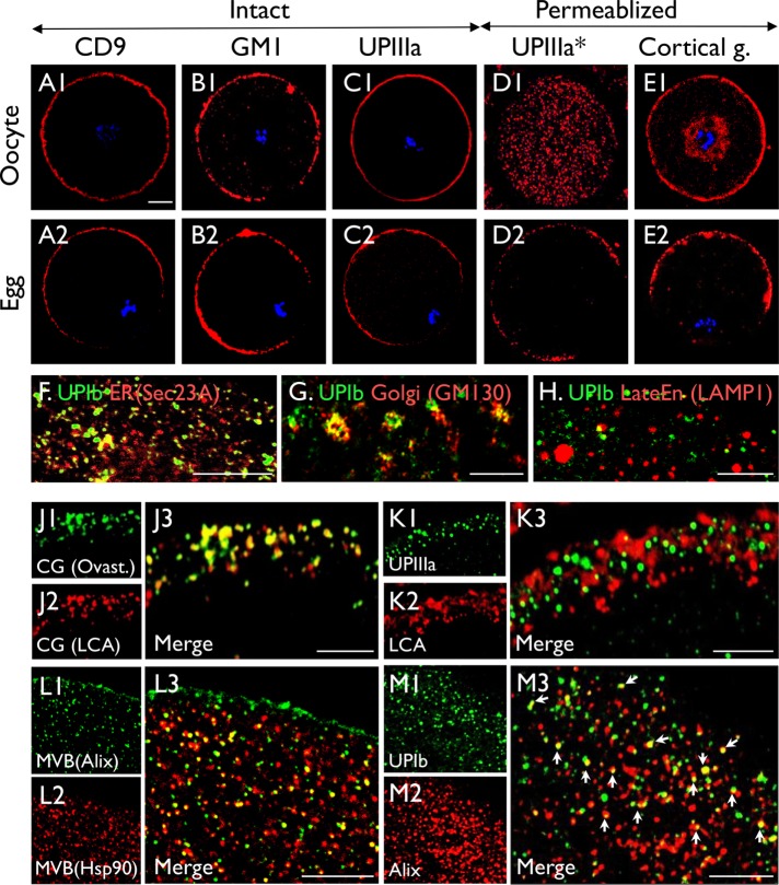
FIGURE 2:
Colocalization of uroplakins with CD9 and CD81 in polarizing mouse oocytes. (A–E) immunofluorescence staining of immature oocytes (A1–E1) and mature eggs (A2–E2) using (A) anti-CD9, (B) Cholera toxin (CTB, recognizing ganglioside GM1, a raft marker); (C, D) anti-uroplakin IIIa (35804); and (E) LCA). The staining was performed using intact oocytes and eggs (A, B, C), Triton X-100–treated samples (E), or paraffin sections (D*). Note the relatively even surface-association of uroplakins and other surface markers in immature oocytes (A1–E1), and their polarization to the microvilli-rich pole on egg maturation (A2–E2). Also note the abundant cytoplasmic UP vesicles in permeabilized oocytes (D1), and their predominant polarized surface-association in mature eggs (C2, D2). (F–M) Oocytes were double-stained using antibodies to (F) UPIb (AU-Ib-2)/Sec23A (ER marker); (G) UPIb (AU-Ib-2)/GM130 (Golgi); (H) UPIb (7472)/LAMP1 (late endosome); (J) ovastacin/LCA (both cortical granule markers; note precise colocalization); (K) UPIIIa (35804)/LCA; (L) Hsp90/Alix (both multivesicular body markers); (M) UPIb (7472)/Alix. Note in M the partial colocalization of UPIb with Alix. Bars equal to 10 µm (A-E) or 5 µm (F–M).

