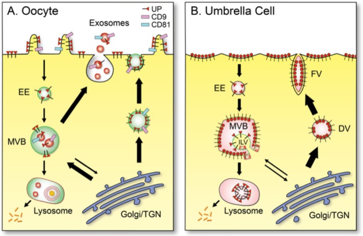
FIGURE 9:
Schematic diagrams showing the distinct patterns of uroplakin trafficking in (A) mouse oocyte/egg and (B) a terminally differentiated umbrella cells of mammalian bladder urothelium. (A) In mouse egg, uroplakins (red triangle luminal domain with a cytoplasmic tail; existing possibly as heterotetramers; see Discussion) are delivered to the cell surface via exocytosis or to exosomes via MVBs. (B) In urothelial umbrella cells, uroplakins are assembled in trans-Golgi network into 16-nm particles (red circles; 16-nm particles), which form growing two-dimensional crystals delivered via DV and FV to the urothelial apical surface, where they form the characteristic urothelial plaques (Wankel et al., 2016). Some of the apical surface-associated uroplakins can be endocytosed into multivesicular vesicles for lysosomal degradation (Vieira et al., 2014). Other abbreviations: EE (early endosome) and TGN (trans-Golgi network). Thickness of the arrows approximates the relative abundance of the pathways in the two cell types.

