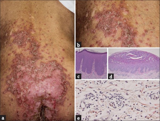
Figure 1.

(a) Deterioration of psoriasis (b) with multiple pustules. (c) Histopathological findings showed regular epidermal proliferation with parakeratosis (H and E, ×100), (d) a subcorneal neutrophilic abscess (H and E, ×40) and (e) infiltration of eosinophils in the upper dermis (Congo-red, ×400)
