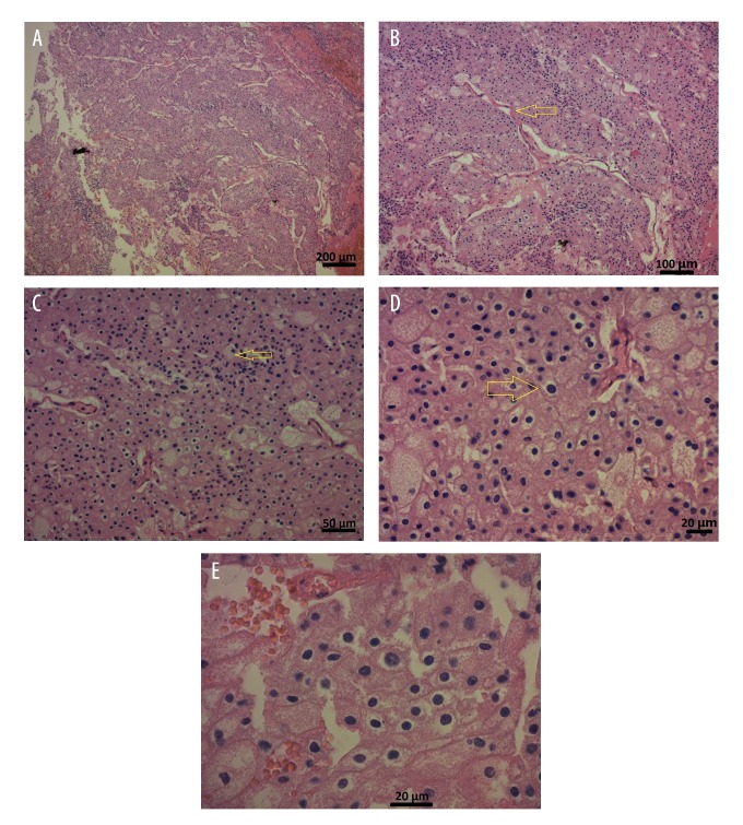
Figure 2.
Photomicrographs of the histopathology of a chromophobe renal cell carcinoma in a 59-year-old man. (A) The photomicrograph shows the characteristic solid growth pattern of a chromophobe renal cell carcinoma. Hematoxylin and eosin (H&E). Magnification ×50. Scale bar, 200 μm. (B) The photomicrograph shows the tumor cells growing in solid nests divided by vascular septae (arrow). H&E. Magnification ×100. Scale bar, 100 μm. (C) The photomicrograph shows that the tumor cells are large and pale with reticulated cytoplasm and perinuclear halos (arrow). H&E. Magnification ×200. Scale bar, 50 μm. (D) The photomicrograph shows large tumor cells with perinuclear halos or translucent zones (arrow). The cytoplasm is pale and flocculent but not clear. H&E. Magnification ×400. Scale bar, 20 μm. (E) The photomicrograph shows that the tumor cells have irregular, wrinkled, and angulated nuclei with perinuclear halos. H&E. Magnification ×630. Scale bar, 20 μm.

