Figure 1.
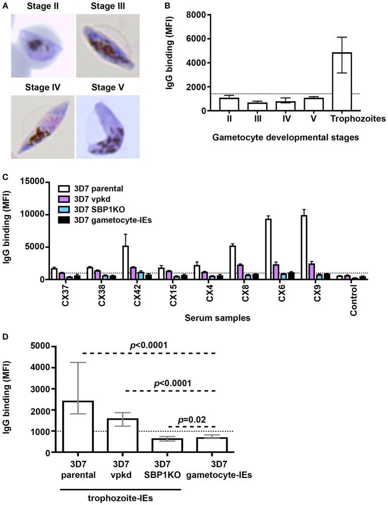
Low levels of naturally-acquired antibodies to the surface of gametocyte-IEs. (A) Giemsa-stained smears confirm the respective gametocyte-IE stages. (B) Total IgG binding to the surface of trophozoite-IEs and gametocyte-IEs was measured at stages II–V of gametocyte development. Samples were from malaria-exposed Kenyan individuals (children n = 11 and adults n = 10). The dotted line represents the antibody positivity threshold (MFI levels greater than mean + 3SD of non-exposed Melbourne controls). IgG binding levels are expressed as geometric mean fluorescence intensity (MFI) for all graphs; assays were performed thrice independently, with samples measured in duplicate (n = 21); bars represent mean and standard deviation. (C) A representative selection of plasma samples tested for antibodies to trophozoite-IEs and gametocyte-IEs. 3D7vpkd and 3D7-SBP1KO are transgenic parasite lines with inhibited PfEMP1 surface expression through the suppression of endogenous var genes (var promoter “knock-down”; vpkd) (21, 26) or genetic deletion of the PfEMP1 trafficking protein (skeleton-binding protein 1 “knock-out”; SBP1KO) (22, 27, 28); these were used at the asexual mature trophozoite stage. Samples were from malaria-exposed Kenyan individuals (CX; children n = 11 and adults n = 10) and non-exposed Melbourne residents (Control). IgG binding to gametocyte-IEs was substantially lower in all individuals compared to IgG binding to trophozoite-IEs. There was minimal background reactivity observed among sera from Melbourne residents; the dotted line represents the antibody positivity threshold. Assays were performed thrice independently; bars represent mean and range of samples tested in duplicate. (D) IgG binding to the surface of stage V 3D7 gametocyte-IEs was substantially lower compared to trophozoite-IEs of 3D7 parental and 3D7vpkd. The difference in IgG binding between gametocyte-IEs and trophozoite-IEs of 3D7 SBP1KO was minimal in our sample set. The dotted line represents the antibody positivity threshold (MFI levels greater than mean + 3SD of non-exposed Melbourne controls). Assays were performed thrice independently; bars represent median and interquartile ranges of samples tested in duplicate (n = 21; children n = 11 and adults n = 10); p-values were calculated using a paired Wilcoxon signed rank test.

