Figure 2.
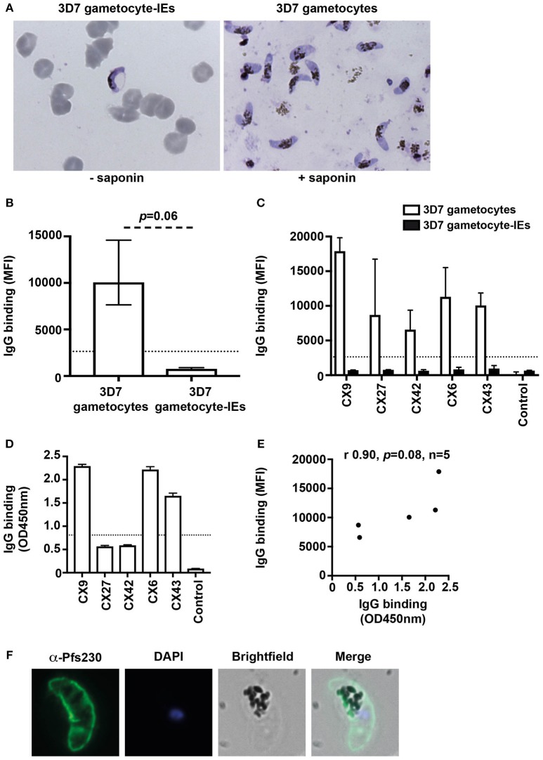
Antibodies recognize the surface of gametocytes in contrast to gametocyte-IEs. (A) Giemsa smears of gametocytes (without the erythrocyte membrane) and intact gametocyte-IEs that were used in antibody assays. (B) IgG binding to 3D7 gametocytes (without the erythrocyte membrane) was markedly higher compared to intact gametocyte-IEs. Assays were performed thrice independently; bars represent median and interquartile ranges of samples tested in duplicates (n = 5; children n = 3, adults n = 2); p-value was calculated using a paired Wilcoxon signed rank test. The dotted line represents the antibody positivity threshold (MFI levels greater than mean + 3SD of non-exposed Melbourne controls). (C) A representative selection of plasma samples tested for antibodies to 3D7 gametocytes and intact gametocyte-IEs. Samples were from malaria-exposed Kenyan individuals (CX; children n = 3, adults n = 2), and non-exposed Melbourne residents (Control). IgG binding to 3D7 gametocytes was substantially higher in all individuals compared to IgG binding to intact gametocyte-IEs. There was minimal background reactivity observed among sera from Melbourne residents; the dotted line represents the antibody positivity threshold. Assays were performed thrice independently; bars represent mean and range of samples tested in duplicate. (D) A representative selection of plasma samples were tested for total IgG binding to recombinant Pfs230D1M. The same selection of samples measured by flow cytometry (n = 21; children n = 11 and adults n = 10) was used. Antibody levels are expressed in optical density (OD) measured at 450 nm. Assays were performed twice; bars represent mean and range of samples tested in duplicate (n = 21); the dotted line represents the antibody positivity threshold. (E) There was a strong positive (non-significant) correlation between total IgG binding measured by flow cytometry (MFI) to 3D7 gametocytes and by ELISA (OD450 nm) to recombinant Pfs230D1M. Correlations were evaluated using Spearman's rho (r). (F) Immunofluorescence microscopy demonstrates the recognition of the native gametocyte surface by a Pfs230-specific antibody (green). Cells were fixed with 90% acetone and 10% methanol, and DAPI was used to stain nuclear DNA (blue). Representative images are shown.

