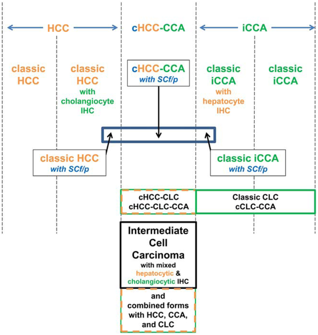
FIG. 1.
Differentiative and histological relationships between PLCs. PLCs show an array of differentiative states from hepatocytic (left) through combined types (middle) to cholangiocytic (right). Thus, there are classic HCCs that are morphologically only hepatocytic, some of which, however, display immunophenotypes, including cholangiocytic marker antigens (e.g., K19). There are also classic iCCAs that are morphologically pure adenocarcinomas, some of which may display immunophenotypes of hepatocytic marker antigens or mRNA. In the middle are the cHCC-CCAs—these are histologically partly morphological HCC and partly morphologically iCCA; immunophenotyping of such lesions can be helpful in confirming the histological impression, but morphology remains the primary criterion. CLC is a separate form of generally lower-grade biliary malignancy. CLC, as indicated, may be found in combination with any of the other forms of PLC in the diagram. Intermediate cell carcinoma is also distinctive: For this tumor, the morphology is neither that of HCC nor that of iCCA, but the mixture of cholangiocytic and hepatocytic features is observed on a cell-by-cell basis on the basis of immunophenotyping. Thus, for this tumor type alone, morphology requires confirmatory immunophenotyping to demonstrate the mixture of differentiation markers.

