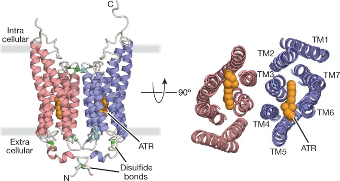
Fig. 1. Overall structure of GtACR1.

Crystal structure of the GtACR1 dimer, viewed parallel to the membrane (left) and from the extracellular side (right). Disulfide bonds are shown using a stick model (green), and ATR (orange) is depicted by a sphere model.
