
Figure 3.
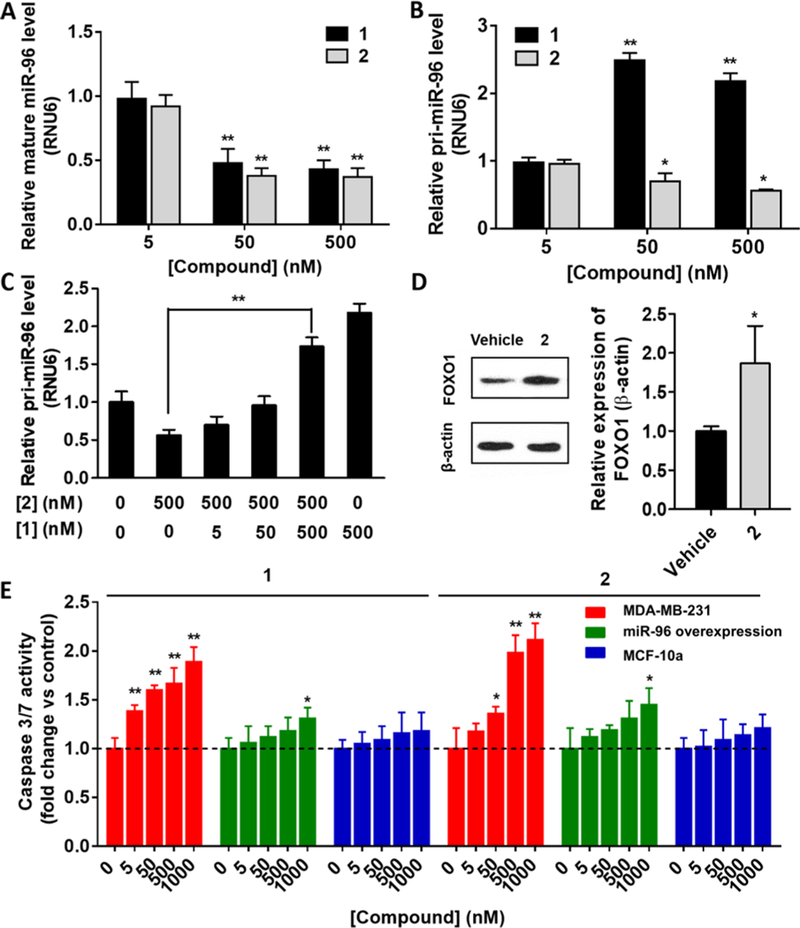
Studying the effect of 1 and 2 on mature miR-96 and pri-miR-96 levels and on miRNA-mediated biology. (A) Effects of 1 and 2 on mature miR-96 levels in MDA-MB-231 TNBC cells, as determined by RT-qPCR. (B) Effects of 1 and 2 on pri-miR-96 levels in MDA-MB-231 cells. As expected based on their modes of action, 1 (simple binding) increased pri-miR-96 levels, while 2 (cleavage) reduced them. (C) Co-addition of increasing concentrations of 1 (5 to 500 nM) and a constant concentration of 2 (500 nM) to MDA-MB-231 cells increased levels of pri-miR-96, diminishing the cleaving capacity of 2 as expected. (D) Effect of 2 on expression of FOXO1 protein, a direct target of miR-96, as determined by Western blot. (E) Effect of 1 or 2 on apoptosis in MDA-MB-231 cells (red), MDA-MB-231 cells that overexpress pri-miR-96 via a plasmid (green), and in MCF-10a healthy breast epithelial cells (blue), as determined by Caspase assays. Data are expressed as mean ± SEM (n > 3). *p < 0.05, **p < 0.01, as measured by a two-tailed Student t test by comparison to untreated cells.