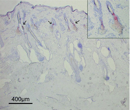
Figure 3.

A section of bovine skin stained with Oil Red O to visualize adipocytes. All the dermis appeared negative to this staining, attesting to the absence of adipose cells. Oil Red O staining revealed the lipids in the inner part and the excretory duct of the sebaceous glands (arrows) localized in the upper part of the dermis. In the inset, a high-power magnification of two sebaceous glands positive to Oil Red O staining is shown. The gland ducts opening in the hair canals can be observed.
