Figure 4.

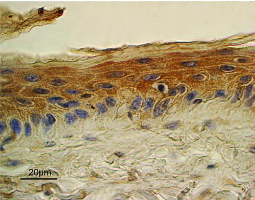
Lep expression in the epidermis. A diffuse staining was mainly observed in the suprabasal cell layers, compared to basal cells. ABC immunohistochemical staining; nuclei are counterstained with Haematoxylin.

Lep expression in the epidermis. A diffuse staining was mainly observed in the suprabasal cell layers, compared to basal cells. ABC immunohistochemical staining; nuclei are counterstained with Haematoxylin.