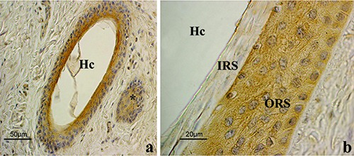
Figure 5.

Lep immunohistochemistry in anagen HFs. a) Low magnification of an oblique section done at the level of the isthmus. Immunostaining involves the ORS cells. The positive structure (*) on the right is the wall of another HF. b) High power magnification of the isthmic region. Cytoplasmic staining of the ORS is clearly shown while the IRS appeared negative; ABC immunohistochemical staining; nuclei are counterstained with Haematoxylin. ORS, outer root sheath; IRS, inner root sheath; Hc, hair canal.
