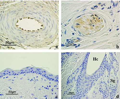
Figure 8.

Positive and negative controls of the immunoreactions. a) Endothelial cells of a vessel in the dermis appeared positive to Lep. b) A nervous fiber in the dermis appeared positive for LepR. c) Negative control for Lep showing the epidermis and (d) for LepR showing a HF and sebaceous glands. The immunostaining is absent in all these structures. Hc, hair canal; Sg, sebaceous glands. All sections are counterstained with Haematoxylin.
