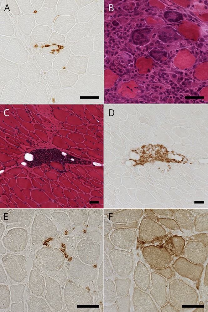
Figure 2. Histopathologic findings of patients with both inflammatory myopathy and myasthenia gravis.

(A) Immunohistochemical staining for CD8 showing CD8-positive cells invading non-necrotic muscle fibers in patient 1. (B) Hematoxylin-eosin (HE) staining showing granulomatous lesion with multinucleated giant cells in patient 7. (C and D) Serial sections with HE staining and immunohistochemical staining for CD20 showing perimysial aggregation of CD20-positive cells in patient 8. (E and F) Serial sections with immunohistochemical staining for PD-1 and PD-L1 showing endomysial PD-1–positive cells and PD-L1 upregulation on non-necrotic fibers in patient 9. Scale bar = 50 μm. PD-1 = programmed cell death 1; PD-L1 = programmed cell death ligand 1.
