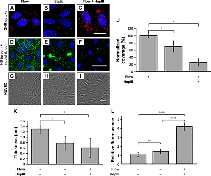
Figure 6.
GCX expression and GNR uptake in HUVECs under different enzyme and flow conditions.
Notes: (A–I) Images of HUVEC cultures conditioned by 16 hours of 12 dynes/cm2 shear stress flow, absence of flow, or flow combined with GCX damaged by 1.25×10⁻⁶ IU/mL HepIII enzyme to degrade HS. (A–C) These conditions were followed by 4 hours of incubation with GNRs. The nanoparticles are shown in red and HUVEC nuclei are shown in blue. Scale bar =20 µm. (D–F) Confocal images of HUVEC cultures stained for the HS component of GCX, for all three conditions. Scale bar =50 µm. (G–I) Phase contrast images show healthy HUVEC morphology, indicating HUVEC compatibility with GNR treatments. Scale bar =50 µm. (J–L) GCX measurements of GCX coverage (J), GCX thickness (K), and corresponding GNR uptake (L). Data shown are mean ± SEM. *P<0.05, **P<0.01, ****P<0.0001, and 3,N,5.
Abbreviations: GCX, glycocalyx; GNRs, gold nanorods; HUVEC, human umbilical vein endothelial cell.

