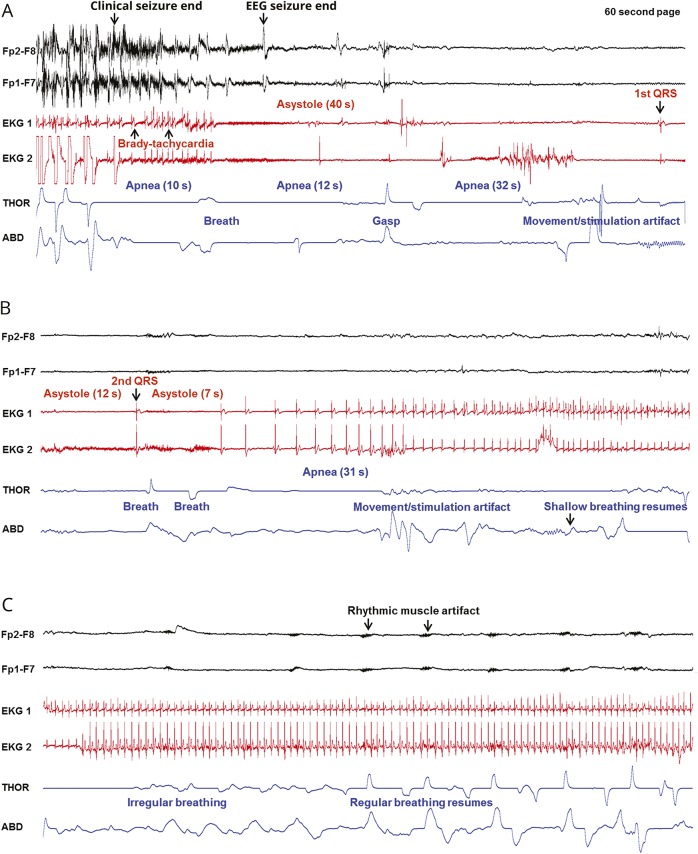
Figure 2. Sequence of EEG, ECG, and breathing changes in the near-SUDEP event of patient 1.
(A–C) Clinical seizure end and onset of the postictal convulsive phase are shown in 3 consecutive 60-second pages. Two channels of the EEG recording are displayed, along with 2 ECG channels and thoracic (THOR) and abdominal (ABD) belts. (A) After clinical seizure end, apnea is noted, accompanied by bradytachycardia that progresses into asystole. Three apneic periods are seen. The first QRS complex is seen at the end of the page. (B) Apnea and asystole continue from panel A until a second QRS complex and 2 breaths, followed by further apnea and asystole. Regular cardiac rhythm is progressively restored, although apnea persists until several seconds later. (C) Cardiac rhythm is re-established, and breathing excursions become more regular and increase in amplitude. Rhythmic muscle artifact becomes more evident on EEG. SUDEP = sudden unexpected death in epilepsy.

