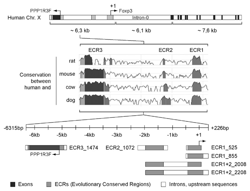
Fig. 1.
Schematic overview of the human Foxp3 locus, the identified ECRs and the amplified promoter fragments. The upper section of the figure shows the organization of the FOXP3 gene on chromosome X. The gene PPP1R3F (NM_033215) is located upstream. The three ECRs on the upstream region of human Foxp3 (indicated in gray) were identified based on the DNA sequence similarity between the human, rat, mouse, cow and dog FOXP3 loci using the bioinformatics program ‘ECR-Browser’ (database May’04/hg17; human Chr. X: 48883883–48863781; conservation: minimum length 100 base pairs, 85% identity). The cloned promoter fragments containing the identified ECRs are illustrated in the lower section. The black areas correspond to exons.

