
Figure 6.
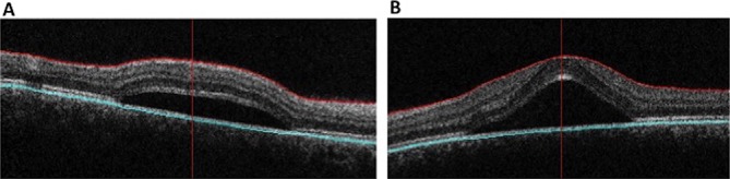
Automated OCT of the right (A) and left (B) maculae in a 30-year-old woman complaining of a blurry spot in the left eye. Visual acuity was 20/20 in the right eye and 20/50 in the left eye. OCT revealed subfoveal collections of fluid in both eyes. The patient was on chronic oral prednisone therapy for systemic lupus erythematous and was diagnosed with bilateral central serous chorioretinopathy. OCT, optical coherence tomography.