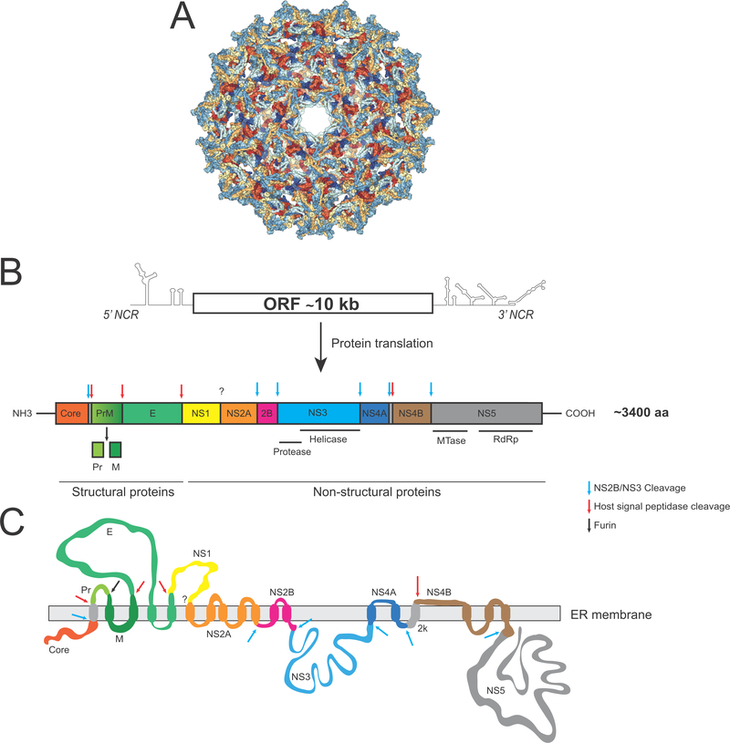
Figure 2. YFV genome organization.
A. Cryo-EM representation of an immature YFV particle (PDB 1NA4). B. Schematic representation of YFV viral RNA and polyprotein. Each viral protein is represented using a distinct color. Arrows indicate cleavage sites in the polyprotein that are processed by proteases of cellular (red or black arrow) or viral (blue arrow) origin. C. Schematic representation of the YFV polyprotein anchored into the endoplasmic reticulum (ER) membrane following translation.

