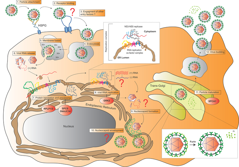
Figure 3. Schematic representation of YFV life cycle.
Key steps of the YFV replication life cycle are displayed from 1 to 11. The few identified host factors regulating some of these steps are shown in red circles (see section 3 for description). The viral RNA replication step and the PrM-E maturation step are enlarged in white boxes. Major gaps in our understanding of specific steps of the life cycle are highlighted by red question marks.

