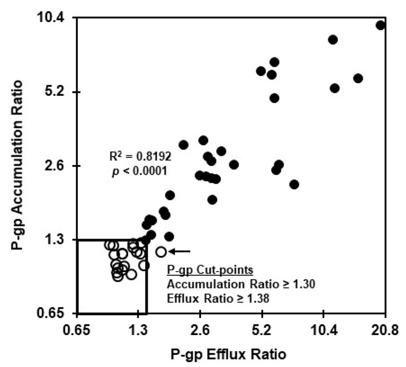
Figure. 2.
Comparison of P-gp ratios generated with the accumulation and efflux bioassays. Fifty AML specimens were tested in the presence and absence of zosuquidar in the DiOC2 accumulation and efflux bioassays as described in the legend to figure 1. P-gp accumulation and efflux ratios were calculated as described for Table 1. Samples that tested P-gp-negative using the accumulation bioassay are indicated by open symbols, while P-gp-positive samples are denoted as solid symbols. The accumulation and efflux bioassays were comparably effective at differentiating P-gp-positive vs. –negative AML specimens.

