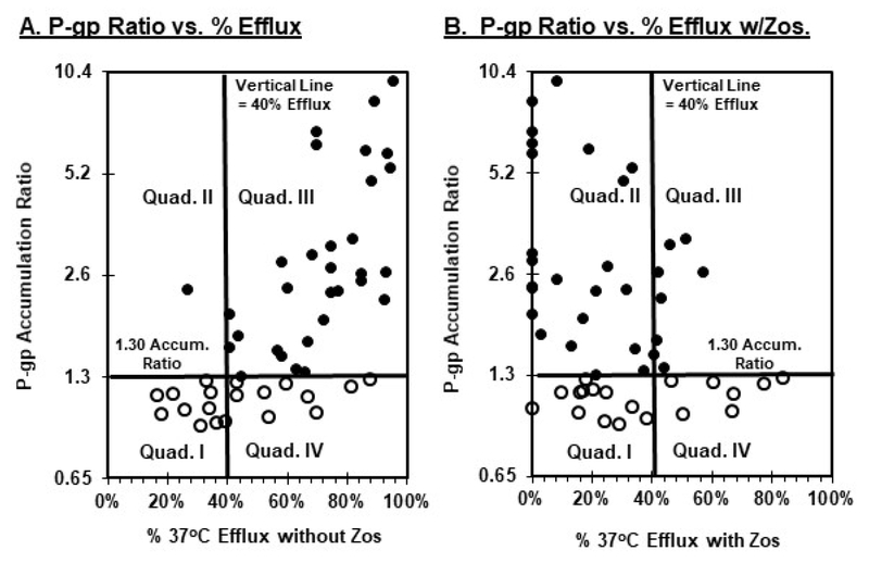
Figure 3.
Identification of independent and concomitant P-gp and non-P-gp MDR function by AML blasts with the accumulation and efflux bioassays. Bioassay results were obtained as described in the legend to figure 1. Solid symbols identify specimens identified as P-gp-positive, while open symbols denote specimens lacking P-gp function. Panel A illustrates P-gp accumulation ratios (x-axis) vs. percent 37°C efflux in the absence of zosuquidar (y-axis). Panel B shows P-gp accumulation ratios (x-axis) vs. percent 37°C efflux in the presence of zosuquidar (y-axis).

