
Figure 5. Transitional macrophages expressing CX3CR1, MHCII, and SiglecF localize to sites of fibroblast accumulation after lung injury.
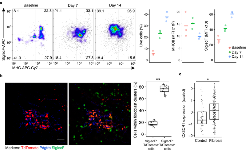
a, Flow cytometry of TdTomato+ lung cells in Cx3cr1-CreERT2 / Rosa26-loxp-STOP-loxp-TdTomato mice with tamoxifen administration before and after injury followed by flow cytometry, with representative pseudocolor plots and values plotted for TdTomato+ cells at three different time points (n=3 biologically independent mice per group, mean is marked). b, Lung immunofluorescence at 14 days after injury in Cx3cr1-CreERT2 / Rosa26-loxp-STOP-loxp-TdTomato mice treated as in a (representative images are shown; quantitation is for n=3 biologically independent mice, 5 images per mouse). Scale bar, 50 μm. c, Expression of CX3CR1 in bulk RNA-seq samples from patient lung biopsy specimens32. Box plot center lines are median, box limits are upper and lower quartiles, and whiskers denote the largest and smallest values no more than 1.5 times the interquartile range from the limits. Wilcoxon test 2-sided p-values are presented. * p <0.001; ** p < 0.0001.