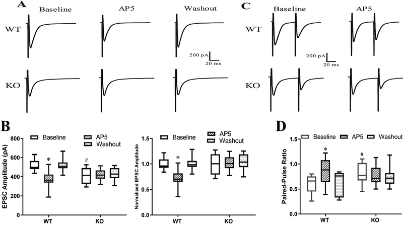
Figure 8. Ablation of α2δ−1 abolishes paclitaxel-induced activation of presynaptic NMDARs at primary afferent terminals in mice.
(A,B). Original recording traces (A) and box-and-whisker plots (B) shows the effect of bath application of 50 μM AP5 on evoked monosynaptic EPSCs of lamina II neurons from wild-type (WT, n = 13 neurons from 5 mice) and α2δ−1 knockout (KO, n = 11 neurons from 5 mice) mice treated with paclitaxel. In B (right panel), values are normalized to the respective baselines. (C,D). Original recording traces (C) and box-and-whisker plots (D) shows the effect of bath application of AP5 on the paired-pulse ratio (PPR) of lamina II neurons from WT (n = 13 neurons from 5 mice) and α2δ−1 KO (n = 10 neurons from 5 mice) mice treated with paclitaxel. *P < 0.05 compared with the respective baseline control (one-way ANOVA followed by Dunnett’s post hoc test). #P < 0.05 compared with the baseline level in WT mice (one-way ANOVA followed by Tukey’s post hoc test).

