
Figure 10.
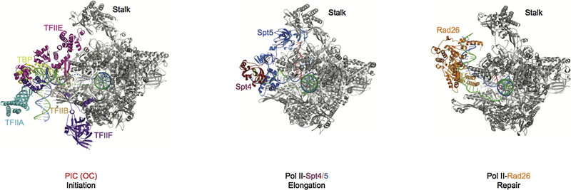
Comparison of three stages of Pol II complexes. The initiation open complex structure is shown on the left, PDB ID: 5FYW; The transcription elongation complex is shown in the middle, PDB ID: 5OIK; The transcription repair initiation complex is shown on the right, PDB ID: 5VVR. In the three different stages, TFIIE, Spt4/5, and CSB bind to similar positions on Pol II. Pol II is in gray and other factors are highlighted and labeled as indicated. Template and non-template DNA, and RNA are shown in blue, green, and red, respectively.