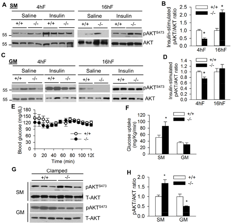
Fig. 3. Fasting-dependent insulin signaling in skeletal muscle of Bscl2−/− mice.

(A) Representative western blot of saline (Sal) and insulin-stimulated AKT Ser473 phosphorylation and (B) quantification of the ratio of insulin-stimulated pAKT/AKT in soleus muscle (SM). (C) Representative western blot of saline (Sal) and insulin-stimulated AKT Ser473 phosphorylation and (D) quantification of the ratio of insulin-stimulated pAKT/AKT in gastrocnemius muscle (GM). 10-13 week old male mice were injected with saline or insulin at 1 unit/kg BW by ip after 4 h fast (4hF) or overnight 16 h fast (16hF). Tissues were harvested after 15 mins. n=4/group. (E) Blood glucose during a 2 h hyperinsulinemic-euglycemic clamp and (F) glucose uptake in soleus muscle (SM) and gastrocnemius muscle (GM) of overnight (16h) fasted Bscl2+/+ (+/+) and Bscl2−/− (−/−) mice after hyperinsulinemic-euglycemic clamp. n=6/group. (G) Insulin mediated AKT Ser473 phosphorylation and (H) ratios of pAKT/AKT in SM and GM under clamp. Three representative animals were shown for each group (n=6 total). *: p< 0.05 vs Bscl2+/+ mice.
