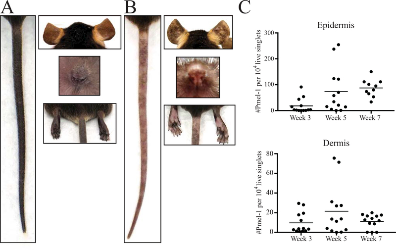
Figure 2:
Vitiligo development in SCF mice. A) Images of the tail, ears, nose and rear footpad of an SCF mouse without vitiligo. B) Images revealing spots of depigmentation on the tail, ears, nose and rear footpads of an SCF mouse 7 weeks post vitiligo induction. C) The number of infiltrating Pmel-1 (normalized to live singlets) in the epidermis and dermis during vitiligo progression.

