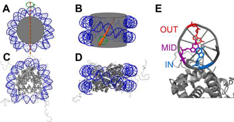
Fig. 1.
Representations of the NCP. (A) Top view and (B) side view of the NCP represented as a spool-and-thread cartoon. The DNA is colored blue and the histone octamer core is gray. The dyad axis is indicated by a dashed orange line in (A) and an orange rod coming out of the plane of (B). The pseudosymmetry of the histone octamer about the dyad axis is indicated by the green arrow. In (C) and (D) the histone proteins are shown as ribbon diagrams and images are merged crystal structures of an NCP containing the Widom 601 DNA with a histone octamer containing N-terminal tail regions (Protein Data Bank entries 3lz0 and 1kx5, respectively). (E) Rotational positions of nucleobases relative to the histone octamer core (only one DNA strand is shown for simplicity). Outward (red), midway (purple), and inward (blue) bases are indicated.

