Figure 1.

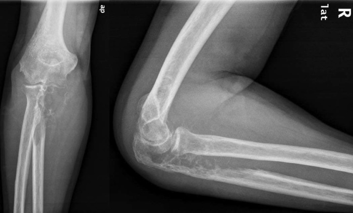
(Left) Anteroposterior (ap) and (right) lateral (lat) x-ray images of the right elbow show an osteolytic lesion of the proximal ulna involving the entire ulnohumeral articulation with soap bubble appearance.

(Left) Anteroposterior (ap) and (right) lateral (lat) x-ray images of the right elbow show an osteolytic lesion of the proximal ulna involving the entire ulnohumeral articulation with soap bubble appearance.