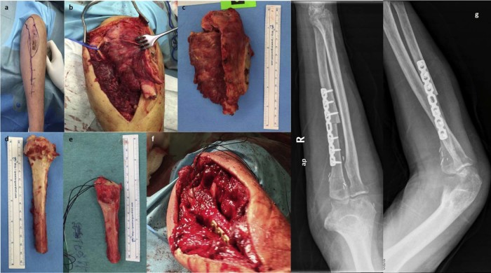
Figure 2.
Intraoperative photographs: (a) the posterior approach used, including the site of the previous biopsy, has been drawn on the arm, (b and c) excision of the tumor using microsurgical technique with protection of the ulnar nerve (arrow), (d and e) the autogenous free fibular graft before and after its preparation with transosseous sutures for triceps reattachment and proper trimming to accommodate the exact length of the bone defect, and (f) the graft is shown in place. (g) Postoperative (left) anteroposterior (ap) and (right) lateral (lat) x-ray images show the reconstruction.

