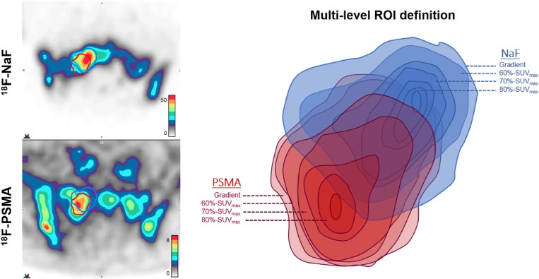
Figure 4. Illustrative example of multi-level segmentation by tracer activity.
Gradient-based segmentation (outermost level) demonstrates moderate spatial concordance of ROI volumes in right sacral bone lesion of castration-resistant patient (serum PSA 4379 ng/ml) imaged with 18F-PSMA (DCFBC) and 18F-NaF. For each radiotracer, additional ROIs are then derived from voxels within gradient-based volumes that fall within 60% of SUVmax, 70% of SUVmax, and 80% of SUVmax. Thus, each ROI is incrementally smaller in volume and higher in uptake relative to SUVmax of each tracer within each bone lesion. The example provided demonstrates spatially discordant regions of highest uptake, with decreasing areas of ROI overlap at increasing levels of relative tracer intensity.

