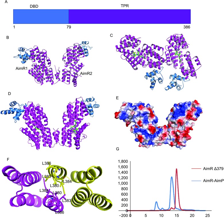
Figure 1.

Overall structures of apo AimR and AimR-AimP complex. (A) Domain organization of AimR. (B) Structure of apo AimR. Two AimR molecules are found in one asymmetric cell unit. The AimR molecules are shown in cartoon, with colors depicting DBD and TPR domains as in Fig. 1A. (C) Structure of AimR-AimP complex in one symmetric cell unit. (D) The homodimer of AimR-AimP. AimRs are colored as in Fig. 1B, whereas AimP molecules are presented as yellow sticks. (E) Surface features of AimR homodimer by electrostatic potential (red, negative; white, neutral; blue, positive). (F) An overhead view of the leucine residues of C-terminal capping helix that form AimR dimerization interface. (G) Analytical size-exclusion chromatography of AimRΔ379 and wild-type AimR-AimP with Superdex 200 increase (GE Healthcare). Before the assay, AimP was incubated with AimR at 10:1 molar ratio for 30 min. Wild-type AimR-AimP was eluted at 13.5 mL (blue), whereas AimRΔ379 was eluted at 14.9 mL (red)
