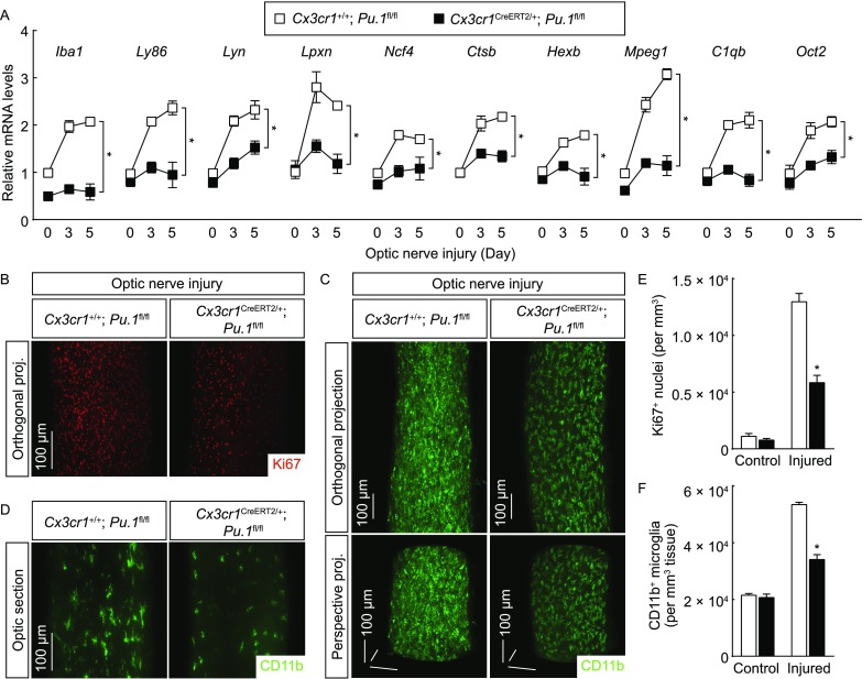
Figure 7.

Post-developmental PU.1 is essential for microglial activation. Cx3cr1+/+; Pu.1fl/fl vs. Cx3cr1CreERT2/+; Pu.1fl/fl mice were treated with 4-hydroxytamoxifen to induce the Cre-recombinase activity and then subjected to optic nerve injury. (A) Expression levels of the indicated genes in the optic nerves were examined by qPCR analysis. n = 4, mean ± SEM, *P < 0.01 (ANOVA test). (B–F) The control and injured nerves were processed for the whole-tissue immunolabeling of Ki67 (B and E) or CD11b (C, D and F) and imaged on the lightsheet microscope. (B) Representative orthogonal 3D projections of the Ki67-immunolabeled injured nerves. (E) Density of Ki67+ nuclei. n = 3, mean ± SEM, *P < 0.01 (ANOVA test). (C) Representative orthogonal (upper panels) or perspective (lower panels) 3D projections of the CD11b-immunolabeled injured nerves. (D) Representative optical sections of the CD11b-immunolabeled injured nerves. (F) Density of CD11b+ microglia. n = 4, mean ± SEM, *P < 0.01 (ANOVA test)
