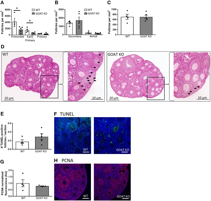
Figure 1.
Effects of GOAT deletion on ovarian follicles in juvenile mice. (A) Small follicle; (B) Large follicle; and (C) Atretic follicle counts in the ovary of juvenile GOAT KO and WT mice. Follicle numbers are expressed per mm3 of ovarian tissue. Panel (D) shows morphological representation of H and E stained juvenile WT and GOAT KO ovary. Black arrows in inserts at higher magnification indicate small follicles (i.e., primordial, early primary and primary) in ovarian cortical region. Scale bars = 20 μm in low and 10 μm in high magnification images. (E) Number of TUNEL-positive follicles in the ovary of juvenile GOAT KO and WT mice. (F) Representative images of TUNEL-positive staining (green) in the juvenile ovary. (G) Normalized mean fluorescence intensity of proliferating cell nuclear antigen (PCNA) staining in juvenile GOAT KO and WT ovaries. (H) Representative images of PCNA staining (red) in the juvenile ovary. Scale bars = 50 μm. Data are expressed as mean ± SEM. *p < 0.05.

