Figure 1.
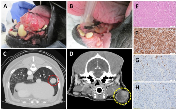
Canine OMM (A) pre- and (B) post- palliative radiation therapy; note the achievement of strong partial response of primary tumor (courtesy of Dr. Michael Kent, UC Davis). Computed tomography of (C) distant pulmonary (red) and (D) regional lymph node metastases (yellow) of OMM origin; demonstrating the potential for reproducible and quantitative volumetric assessments for documentation of abscopal activities. Panel (E–H; top to bottom) represents the histologic and immunobiologic assessment of a regional lymph node effaced with amelanotic melanoma by H&E, PD-L1, CD3+ tumor-infiltrating lymphocytes, and regulatory T cells (courtesy of Dr. Jonathan Samuelson, UIUC). Magnification 400x.

