Figure 5.

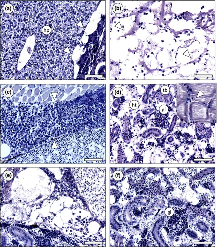
Zebrafish histology (Davidson, H&E). (a) Exemplificative section across the abdominal cavity of a control animal, highlighting hepatic tissue (hp) and interorgan adipose tissue (arrowheads), both devoid of any signs of infection or other pathological features. (b) Section of an animal inoculated with the GAP58 strain, exhibiting severe infection in the interorgan adipose tissue, leading to tissue liquefaction. Cocci can be seen in small clumps of chains, most of which inside the remnants of macrophages (arrowheads). Note that most of the adipose material has been washed‐off during sample processing, leading to potential under evaluation of infection. (c) Massive exudate holding defense cells and bacteria (between arrowheads) in the abdomen of a fish inoculated with the VSD23 strain. The alteration is formed between skeletal muscle and major visceral blood vessel. (d) Severely infected body (trunk) kidney of a zebrafish injected with VSD24 strain. The hematopoietic tissue (ht) shows liquefying necrosis, cocci and macrophage aggregates. Kidney tubules (tb) were obviously less affected but glomeruli (gl) were severely infected as well. Inset: Cocci between fascicles of skeletal muscle, with leukocytes infiltrating adjacently. (e) Section across the abdominal cavity of a fish inoculated with the VSD21 strain, showing severe infection in the interorgan adipose tissue and adjacent kidney. (f) Section of a zebrafish treated with the same strain as previous, revealing acute kidney infection affecting haematopoietic tissue and glomeruli, similarly to panel D. Scale bars: 50 μm
