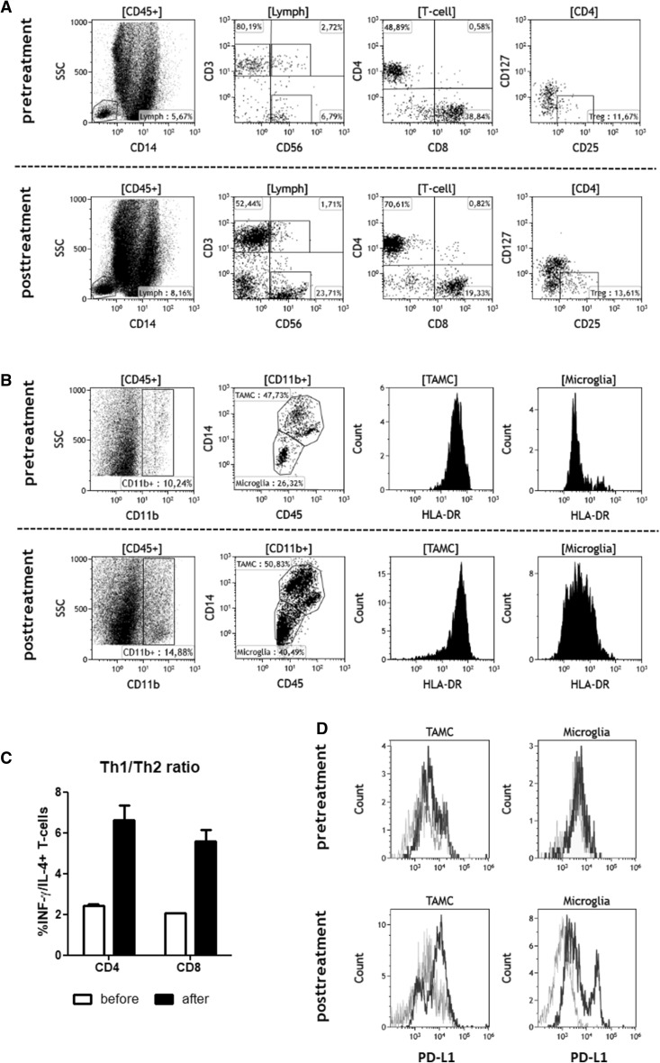
Fig. 3.
Multiparameter flow cytometry. Freshly isolated peripheral blood mononuclear cells (PBMCs) and tumor cell suspensions prepared from resected tumor material were stained using a panel of directly labelled mAbs to detect different T-cell and myeloid cell subpopulations. Gating strategies are described in detail in “Material and Methods”. a Pre- and posttreatment analysis of lymphocyte subpopulations, mainly CD3+, CD4+ and CD8+ T-cells, CD3−CD56+ NK cells and CD4+CD25highCD127low regulatory T-cells. b Pre- and posttreatment analysis of CD45+CD11b+ myeloid cell subsets including HLA-DR expression on tumor-associated CD45++/+++CD14high myeloid cells (TAMC) and CD45+/++CD14low microglia. c Tumor cell suspensions were stimulated with PMA/Ionomycin, and CD4+ and CD8+ CD45RA− memory T cells were analysed for the expression of INF-γ and IL-4 by intracellular FACS staining. Th1/Th2 ratios were calculated by dividing the proportion of IFN-γ positive T-cells by the proportion of IL-4 positive cells. d Pre- and posttreatment expression of PD-L1 on TAMC and microglia (black line = PD-L1; grey line = isotype control)

