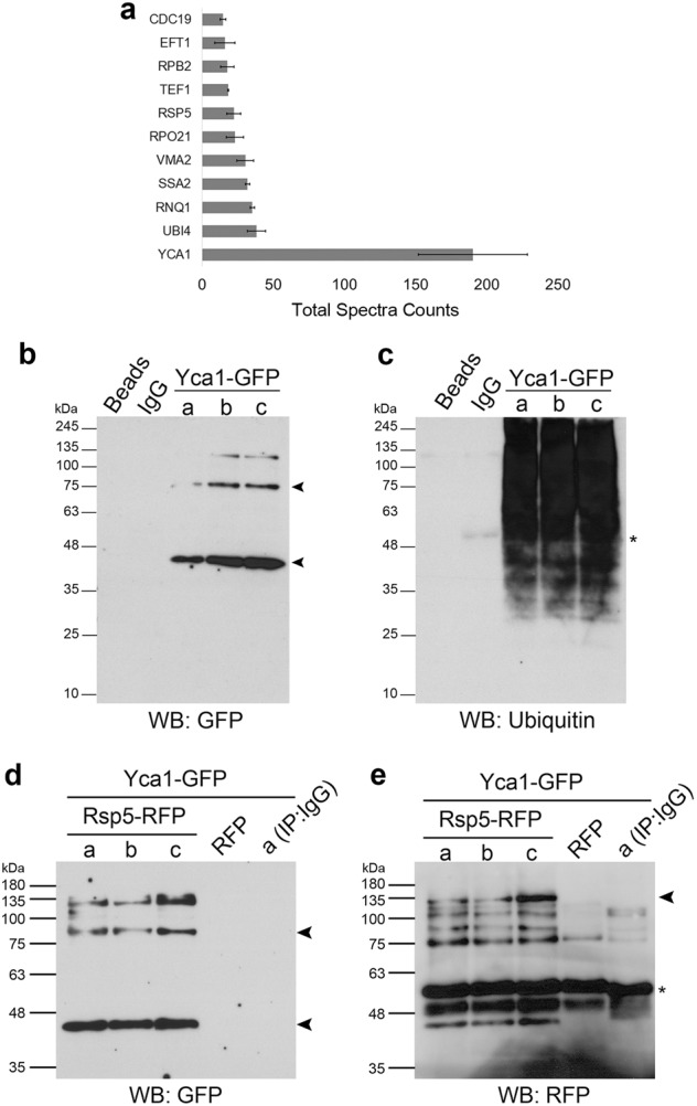
Fig. 1. Yca1 interacts with components of the ubiquitin pathway.

a LC-MS/MS analysis of Yca1-RFP pulldown depicted ubiquitin (Ubi4) and the E3 ligase Rsp5 in high abundance (Total Spectra Counts). Data represented as mean ± SEM. n = 3. b Yca1-GFP cells were used to isolate ubiquitinated proteins which was further probed with anti-GFP antibody to detect the presence of Yca1-GFP. The arrows highlight the full length and processed forms of Yca1-GFP. Predicted size of full length Yca1-GFP is 76 kDa and processed form in 37 kDa. Protein G beads and anti-IgG antibody conjugated beads were used as controls. Lowercase letters denote independent replicates. n = 3. c Extracts in b were probed with anti-ubiquitin antibody to detect the presence of the bait. The asterisk (*) highlights the presence of IgG. d Rsp5-RFP was expressed from a plasmid in the Yca1-GFP strain and assessed for interaction between Yca1 and Rsp5 via anti-RFP immunoprecipitation followed by anti-GFP immunoblotting. Lowercase letters denote replicate samples. The arrows highlight the full length and processed forms of Yca1-GFP. The empty vector transformed Yca1-GFP cells (RFP) was used as an interaction control. Anti-IgG antibody conjugated beads were used to probe replicate ‘a’ as an additional control. n = 3. e Extracts in d were probed with anti-RFP antibody to detect the presence of the bait Rsp5-RFP (highlighted with an arrow). The estimated size of Rsp5-RFP is 119 kDa. The asterisk highlights the presence of IgG
