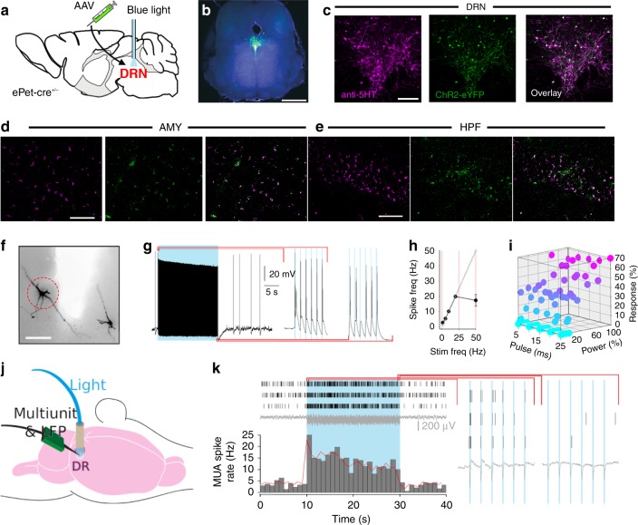
Fig. 1.
Optogenetic targeting of DRN 5-HT neurons. a Infusion of AAV harbouring cre-dependent ChR2-eYFP into the DRN of ePet-cre+/− mice, followed by implantation of a MRI-friendly optic fibre used to deliver blue light. b Whole midbrain slice with Ch2R in green. DAPI (blue) identify all cell nuclei. Scale bar indicates 2.5 mm. c–e Co-immunofluorescence with anti-5-HT (purple) and ChR2-eYFP (green) in the (c) DRN, (d) amygdala (AMY) and (e) hippocampus (HPF). Scale bars indicate 500, 100 and 50 µm, respectively. f Biocytin-filled neurons of the DRN used for in vitro whole-cell patch electrophysiology. Red dashed circle indicates recorded neuron. Scale bar indicates 200 µm. g Response of neuron circled in (f) to all but one pulse of a blue light train. h Frequency response curve from midbrain slices. Linearity stable to 20 Hz. i Strength of response expressed in maximum number of spikes per pulse during 20 Hz photostimulation, as a function of pulse width and laser power. j Schematic of the experimental set-up for paired MUA and LFP recordings in the DRN during photostimulation. k Raster plots of MUA activity in three neighbouring channels (top), LFP (middle) and mean spike histogram (bottom), revealing activation of the DRN 5-HT network during a blue light train (20 Hz, 5 ms pulse width). Expanded to the right are the MUA and LFP for the first and last 6 pulses in the train

